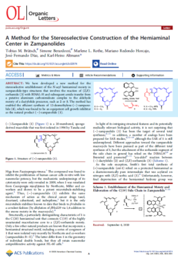
A Method for the Stereoselective Construction of the Hemiaminal Center in Zampanolides
We have developed a new method for the stereoselective establishment of the N-acyl hemiaminal moiety in zampanolide-type structures that involves the reaction of (Z,E)-sorbamide (3) with BINAL-H and subsequent amide transfer from a putative aluminum carboximidoate complex to the aldehyde moiety of a dactylolide precursor, such as 2 or 5. The method has enabled the efficient synthesis of 13-desmethylene-(−)-zampanolide (4), which was found to be an equipotent cell growth inhibitor as the natural product (−)-zampanolide (1).
https://www.tubintrain.eu/wp-content/uploads/2020/10/2020-Brutsch.pdf
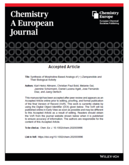
Synthesis of Morpholine-Based Analogs of (−)-Zampanolide and Their Biological Activity
We describe the convergent synthesis of three prototypical examples of a new class of analogs of the complex, cytotoxic marine macrolide (−)-zampanolide, which incorporate an embedded Nsubstituted morpholine moiety in place of the natural tetrahydropyran ring. The final construction of the macrolactone core was based on a high-yielding intramolecular HWE olefination, while the hemiaminallinked side chain was elaborated through a stereoselective, BINAL-Hmediated addition of (Z,E)-sorbamide to a macrocyclic aldehyde precursor. The synthesis of the common functionalized morpholine building block involved two consecutive epoxide openings with tosylamide and the product of the first opening reaction, respectively, as nucleophiles. Of the three morpholino-zampanolides investigated, the N-acetyl and the N-benzoyl derivative both exhibited nanomolar antiproliferative activity, thus being essentially equipotent with the natural product. In contrast, the activity of the N-tosyl derivative was significantly reduced.
https://www.tubintrain.eu/wp-content/uploads/2020/10/2020-Bold.pdf
Microtubule-Targeting Agents:Strategies To HijacktheCytoskeleton
Microtubule-targeting agents(MTAs)such as paclitaxel and the vincaalkaloids are among the most important medical weapons available to combat cancer. MTAs interfere with intracellular transport, inhibit eukaryotic cell proliferation, and promote cell death by suppressing microtubule dynamics. Recent advan-ces in the structural analysis of MTAs have enabled the extensive characterization of their interactions with microtubules and their building block tubulin. We revie where our current knowledge on the molecular mechanisms used by MTAs to hijack the microtubule cytoskeleton,and discuss dual inhibitors that
target both kinases and microtubules. We further formulate some outstanding questions related to MTA structural biology and present possible routes for
future investigations of this fascinating class of antimitotic agents.
The Positive Side of the Alzheimer’s Disease Amyloid Cross-Interactions: The Case of the Aβ 1-42 Peptide with Tau, TTR, CysC, and ApoA1
Alzheimer’s disease (AD) represents a progressive amyloidogenic disorder whose advancement is widely recognized to be connected to amyloid-β peptides and Tau aggregation. However, several other processes likely contribute to the development of AD and some of them might be related to protein-protein interactions. Amyloid aggregates usually contain not only single type of amyloid protein, but also other type of proteins and this phenomenon can be rationally explained by the process of protein cross-seeding and co-assembly. Amyloid cross-interaction is ubiquitous in amyloid fibril formation and so a better knowledge of the amyloid interactome could help to further understand the mechanisms of amyloid related diseases. In this review, we discuss about the cross-interactions of amyloid-β peptides, and in particular Aβ1-42, with other amyloids, which have been presented either as integrated part of Aβ neurotoxicity process (such as Tau) or conversely with a preventive role in AD pathogenesis by directly binding to Aβ (such as transthyretin, cystatin C and apolipoprotein A1). Particularly, we will focus on all the possible therapeutic strategies aiming to rescue the Aβ toxicity by taking inspiration from these proteinprotein interactions.
Combining optogenetics with sensitive FRET imaging to monitor local microtubule manipulations
Optogenetic methods for switching molecular states in cells are increasingly prominent tools in life sciences. Förster Resonance Energy Transfer (FRET)-based sensors can provide quantitative and sensitive readouts of altered cellular biochemistry, e.g. from optogenetics. However, most of the light-inducible domains respond to the same wavelength as is required for excitation of popular CFP/ YFP-based FRET pairs, rendering the techniques incompatible with each other. In order to overcome this limitation, we red-shifted an existing CFP/YFP-based OP18 FRET sensor (COPY) by employing an sYFP2 donor and mScarlet-I acceptor. Their favorable quantum yield and brightness result in a red-shifted FRET pair with an optimized dynamic range, which could be further enhanced by an R125I point mutation that stimulates intramolecular interactions. The new sensor was named ROPY and it visualizes the interaction between the microtubule regulator stathmin/OP18 and free tubulin heterodimers. We show that through phosphorylation of the ROPY sensor, its tubulin sequestering ability can be locally regulated by photo-activatable Rac1 (PARac1), independent of the FRET readout. Together, ROPY and PARac1 provide spatiotemporal control over free tubulin levels. ROPY/ PARac1-based optogenetic regulation of free tubulin levels allowed us to demonstrate that depletion of free tubulin prevents the formation of pioneer microtubules, while local upregulation of tubulin concentration allows localized microtubule extensions to support the lamellipodia.
https://www.nature.com/articles/s41598-020-62874-3.pdf
Direct observation of dynamic protein interactions involving human microtubules using solid-state NMR spectroscopy
Microtubules are important components of the eukaryotic cytoskeleton. Their structural organization is regulated by nucleotide binding and many microtubule-associated proteins (MAPs). While cryo-EM and X-ray crystallography have provided detailed views of interactions between MAPs with the microtubule lattice, little is known about how MAPs and their intrinsically disordered regions interact with the dynamic microtubule surface. NMR carries the potential to directly probe such interactions but so far has been precluded by the low tubulin yield. We present a protocol to produce [13C, 15N]-labeled, functional microtubules (MTs) from human cells for solid-state NMR studies. This approach allowed us to demonstrate that MAPs can differently modulate the fast time-scale dynamics of C-terminal tubulin tails, suggesting distinct interaction modes. Our results pave the way for in-depth NMR studies of protein dynamics involved in MT assembly and their interactions with other cellular components.
Introducing sequential aza-amino acids units induces repeated β-turns and helical conformations in peptides
A major current issue in medicinal chemistry is the design of small peptide analogues resistant to proteolysis and able to adopt preferential conformations, while preserving the selectivity and efficiency of natural peptides. Whereas the introduction of one aza-Gly in peptides has proven numerous biological and structural interest, the conformational effect of sequential aza-Gly or azaamino acids bearing side chains has not been investigated. In this work, experimental NMR and X-ray data together with in silico conformational studies reveal that the introduction of two consecutive aza-amino acids in pseudotripeptides induces the formation of stable hydrogen-bonded β-turn structures. Notably, this stabilization effect relies on the presence of side chains on azaamino acids, as more flexible conformations are observed with aza-Gly residues. Remarkably, a longer aza/aza/α/aza/aza/α pseudohexapeptide containing substituted aza-amino acids adopts repeated β-turns conformations which interconvert with a fully helical structure mimicking a 310 helix.
Tau Biology and Tau-Directed Therapies for Alzheimer’s Disease
Abstract Alzheimer’s disease (AD) is characterised by a progressive loss of cognitive functions. Histopathologically, AD is defined by the presence of extracellular amyloid plaques containing Ab and intracellular neurofibrillary tangles composed of hyperphosphorylated tau proteins. According to the now well-accepted amyloid cascade hypothesis is the Ab pathology the primary driving force of AD pathogenesis, which then induces changes in tau protein leading to a neurodegenerative cascade during the progression of disease. Since many earlier drug trials aiming at preventing Ab pathology failed to demonstrate efficacy, tau and microtubules have come into focus as prominent downstream targets. The article aims to develop the current concept of the involvement of tau in the neurodegenerative triad of synaptic loss, cell death and dendritic simplification. The function of tau as a microtubule associated protein and versatile interaction partner will then be introduced and the rationale and progress of current tau-directed therapy will be discussed in the biological context.
https://www.ncbi.nlm.nih.gov/pmc/articles/PMC4757605/pdf/40265_2015_Article_529.pdf
A Refined Reaction-Diffusion Model of Tau-Microtubule Dynamics and Its Application in FDAP Analysis
ABSTRACT Fluorescence decay after photoactivation (FDAP) and fluorescence recovery after photobleaching (FRAP) are well established approaches for studying the interaction of the microtubule (MT)-associated protein tau with MTs in neuronal cells. Previous interpretations of FDAP/FRAP data have revealed dwell times of tau on MTs in the range of several seconds. However, this is difficult to reconcile with a dwell time recently measured by single-molecule analysis in neuronal processes that was shorter by two orders of magnitude. Questioning the validity of previously used phenomenological interpretations of
FDAP/FRAP data, we have generalized the standard two-state reaction-diffusion equations by 1), accounting for the parallel and discrete arrangement of MTs in cell processes (i.e., homogeneous versus heterogeneous distribution of tau-binding sites); and 2), explicitly considering both active (diffusion upon MTs) and passive (piggybacking upon MTs at rates of slow axonal transport) motion of bound tau. For some idealized cases, analytical solutions were derived. By comparing them with the full numerical solution and Monte Carlo simulations, the respective validity domains were mapped. Interpretation of our FDAP data (from processes of neuronally differentiated PC12 cells) in light of the heterogeneous formalism yielded independent estimates for the association (~2 ms) and dwell (~100 ms) times of tau to/on a single MT rather than in an MT array. The dwell time was shorter by orders of magnitude than that in a previous report where a homogeneous topology of MTs was assumed. We found that the diffusion of bound tau was negligible in vivo, in contrast to an earlier report that tau diffuses along the MT lattice in vitro. Methodologically, our results demonstrate that the heterogeneity of binding sites cannot be ignored when dealing with reaction-diffusion of cytoskeleton-associated proteins. Physiologically, the results reveal the behavior of tau in cellular processes, which is noticeably different from that in vitro.
https://www.ncbi.nlm.nih.gov/pmc/articles/PMC4255205/pdf/main.pdf
Microtubule dynamics and the neurodegenerative triad of Alzheimer's desease: The hidden connection
Alzheimer’s disease (AD) is the most common neurodegenerative disorder and is, on a histopathological level, characterized by the presence of extracellular amyloid plaques composed of the protein fragment Ab, and intracellular neurofibrillary tangles, which contain the microtubule-associated protein tau in a hyperphosphorylated state. In AD defects in microtubule (MT) assembly and organization have also been reported; however, it is unclear whether MT abnormalities have a causal and early role in the disease process or represent a common end point downstream of the neurodegenerative cascade. Recent evidence indicates that microtubule-stabilizing drugs prevent axonopathy in animal models of tauopathies and reverse Ab-induced loss of synaptic connectivity in an ex vivo model of amyloidosis. This could suggest that MT dysfunction connects some of the degenerative events and provides a useful target to simultaneously prevent several neurodegenerative processes in AD. Here, we describe how changes in the structure and dynamics of MTs are involved in the different aspects of the neurodegenerative triad of AD. We discuss evidence that MTs are affected both by tau-dependent and tau-independent mechanisms but appear to be regulated in a distinct way in different neuronal compartments. We argue that modulation of MT
dynamics could be of potential benefit but needs to be precisely controlled in a cell and compartment-specific manner to avoid harmful side effects.
Keywords: cytoskeleton, dendritic spines, microtubule dynamics, tau protein.
https://onlinelibrary.wiley.com/doi/pdf/10.1111/jnc.14011